39 label the structures of a skeletal muscle fiber
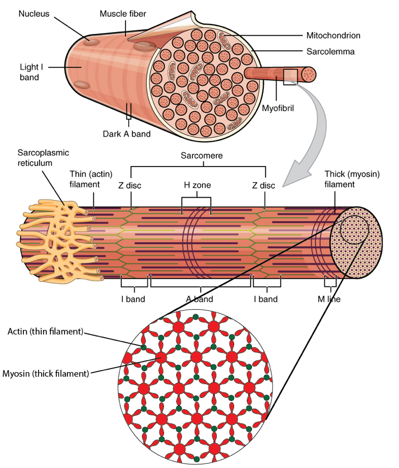
Skeletal Muscle: Structure and Function - ENPAB muscle and their functions. ➤ Draw and label the structures that characterize a skeletal muscle fiber's striated appearance under.23 pages Skeletal Muscle Fiber Structure and Function - Open Textbooks for Hong Kong The orderly arrangement of the proteins in each unit, shown as red and blue lines, gives the cell its striated appearance. The striated appearance of skeletal muscle tissue is a result of repeating bands of the proteins actin and myosin that occur along the length of myofibrils. Myofibrils are composed of smaller structures called myofilaments.
Structures of the Skeletal Muscle Fiber Flashcards | Quizlet -Muscle fibers are filled with threads called myofibrils separated by SR (sarcoplasmic reticulum) -Myofilaments (thick & thin filaments) are the contractile proteins of muscle that actually cause muscles to contract. Sarcoplasmic Reticulum (SR) -Stores Ca+2 in a relaxed muscle -Release of Ca+2 triggers muscle contraction Filaments and the Sarcomere

Label the structures of a skeletal muscle fiber
SKELETAL MUSCLE ORGANIZATION During muscle contraction, the myosin heads link the thick and thin myofilaments together, forming cross bridges that cause the thick and thin myofilaments to slide over each other, resulting in shortening of each sarcomere, each skeletal muscle fiber, and the muscle as a whole-much like the two parts of an extension ladder slide over each other. To summarize, in … General Anatomy of Skeletal Muscle Fibers - GetBodySmart Internal Anatomy of Skeletal Muscle Fibers. An interactive quiz about the internal anatomy of skeletal muscle fibers, featuring illustrations-based multiple choice questions. Skeletal Muscle Fiber Location and Arrangement > Internal Anatomy of Skeletal Muscle Fibers. Subject Areas. Skeletal System; Muscular System; Label the Skeletal Muscle Fiber Quiz - purposegames.com Label the Skeletal Muscle Fiber. ... 11 Tries. 11 Last Played. 28 Apr, 2022 Sound On/Off. From the quiz author. Label Microscopic Anatomy of a Muscle Fiber Remaining 0. Correct 0. Wrong 0. Press play! 0%. 0:00.0. Quit. Again. This game is part of a tournament. You need to be a group member to play the tournament.
Label the structures of a skeletal muscle fiber. Skeletal Muscle Labeling | Biology Quiz - Quizizz Skeletal Muscle Labeling DRAFT. 22 minutes ago. by ggard. Played 0 times. 0. 9th - 10th grade ... Q. Skeletal Muscle contraction is initiated when the _____ sends a message to the muscle cell. ... Muscle cell. Neuron. Gland. None of the above. Tags: Question 30 . SURVEY . 30 seconds . Report an issue . Q. Each skeletal muscle fiber is ... anatomy labeled muscle fiber muscle skeletal fiber structure muscles muscular system fibers human body physiology tendon tissue cell anatomy structures types insertion epimysium origin. Sarcomere . muscle contraction sarcomere anatomy sacromere physiology muscles cell bands unit microscopic basic band dark actin tissue sarcomeres myosin myocyte where Inquizitive CH 23 Flashcards | Quizlet skeletal muscle cardiac muscle smooth muscle. Match each part of the knee with its major function. connect(s) muscle to bone cushions bone to bone connections lubricates and facilitates movement connect(s) bone to bone. Select the image below that best portrays a single, contracted sarcomere. the big three line one. The rib cage is made almost entirely of bone. f. … Skeletal Muscle Fiber | Types, Characteristics & Anatomy - Video ... Structure of Skeletal Muscle Fiber Skeletal muscle fibers are composed of a bundle of thin filaments called myofibrils. Each myofibril is made up of small sections called sarcomeres. Sarcomeres are...
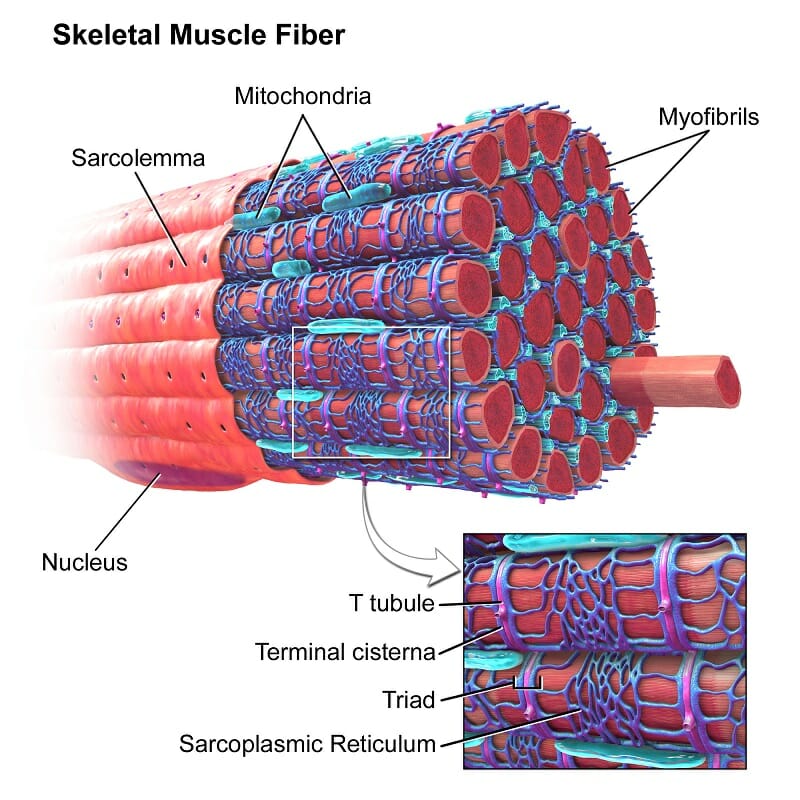
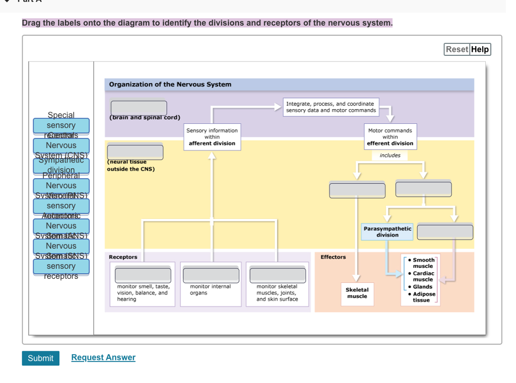
Solved Muscle Cell Label the structures of a skeletal muscle - Chegg Expert Answer 100% (16 ratings) 1) Sarcolemma 2) myofib … View the full answer Transcribed image text: Muscle Cell Label the structures of a skeletal muscle fiber. Nucleus Myofibril Sarcolemma Sarcoplasmic reticulum Openings into T tubules < Prev 3 of 15 !!! Next > Thinkinys - How to write a boty The Good Cre. Internal Anatomy of Skeletal Muscle Fibers - GetBodySmart General Anatomy of Skeletal Muscle Fibers An interactive quiz about the general anatomy of skeletal muscle fibers, featuring illustrations-based multiple choice questions. General Organization of the Nervous System Art-labeling Activity: The Structure of a Skeletal Muscle Fiber Start studying Art-labeling Activity: The Structure of a Skeletal Muscle Fiber. Learn vocabulary, terms, and more with flashcards, games, and other study tools. Nervous System: Explore the Nerves with Interactive Anatomy 02.11.2020 · Efferent neurons (also called motor neurons) carry signals from the gray matter of the CNS through the nerves of the peripheral nervous system to effector cells. The effector may be smooth, cardiac, or skeletal muscle tissue or glandular tissue. The effector then releases a hormone or moves a part of the body to respond to the stimulus.
Skeletal Muscle Fiber Labeling - Printable About this Worksheet. This is a free printable worksheet in PDF format and holds a printable version of the quiz Skeletal Muscle Fiber Labeling.By printing out this quiz and taking it with pen and paper creates for a good variation to only playing it online. Skeletal muscle mass and distribution in 468 men and women … 22.02.2020 · We employed a whole body magnetic resonance imaging protocol to examine the influence of age, gender, body weight, and height on skeletal muscle (SM) mass and distribution in a large and heterogeneous sample of 468 men and women. Men had significantly (P < 0.001) more SM in comparison to women in both absolute terms (33.0 vs. 21.0 kg) and relative to … Anatomy, Skeletal Muscle - StatPearls - NCBI Bookshelf The musculoskeletal system comprises one of the major tissue/organ systems in the body. The three main types of muscle tissue are skeletal, cardiac, and smooth muscle groups.[1][2][3] Skeletal muscle attaches to the bone by tendons, and together they produce all the movements of the body. The skeletal muscle fibers are crossed with a regular pattern of fine red and white lines, giving the ... welcome to Ms. stephens' anatomy and Physiology and … Unit 5: Muscular System Student Learning Goals: I can identify smooth, skeletal, and cardiac muscle tissue under a microscope and state the function of each.; I can identify the component parts of a muscle: fascicle, myofibril, fiber, nucleus of cell, body of muscle.; I can identify the major muscles of the human body.; I can analyze experimental data using the Moving Arm …
Label the Skeletal Muscle Fiber Quiz - purposegames.com Label the Skeletal Muscle Fiber. ... 11 Tries. 11 Last Played. 28 Apr, 2022 Sound On/Off. From the quiz author. Label Microscopic Anatomy of a Muscle Fiber Remaining 0. Correct 0. Wrong 0. Tryk på play! 0%. 0:00.0. Afslut. Igen. This game is part of a tournament. You need to be a group member to play the tournament.
Skeletal Muscle Tissue Anatomy and Structure - Registered Nurse RN Each skeletal muscle is considered an organ, and it's made up of connective tissue layers, muscle fibers, blood vessels, and nerves. Skeletal muscles attach to the bones through tendons or through a direct attachment. As you look at this muscle diagram, you'll notice an outer layer of connective tissue called epimysium.
Label the Skeletal Muscle Fiber Quiz - purposegames.com Label the Skeletal Muscle Fiber. ... Englisch Fragen. 11 Versucht. 11 Zuletzt Gespielt. 28 Apr, 2022 Ton ein / aus. Vom Quizersteller. Label Microscopic Anatomy of a Muscle Fiber Remaining 0. Richtig 0. Wrong 0. Press play! 0%. 0:00.0. Halt. Nochmal. This game is part of a tournament.
Solved Hel Label the structures of a skeletal muscle fiber. - Chegg Question: Hel Label the structures of a skeletal muscle fiber. 4 0.1 points eBook Sarcoplasmic reticulum Nucleus Myofibril Openings into T tubules Sarcolemma Mc Graw Hill < Prey 4 of 20 !!! Next This problem has been solved! See the answer Show transcribed image text Expert Answer 100% (5 ratings)
Skeletal muscle tissue: Histology | Kenhub Special terms are used to describe structures associated with skeletal muscle tissue. Muscle tissue terms often begin with myo-, mys-, or sarco-. The cytoplasm of a muscle cells is referred to as sarcoplasm.The plasma membrane is called the sarcolemma and the endoplasmic reticulum is called the sarcoplasmic reticulum.A muscle fiber may also be referred to as a myofiber.
Structure of Skeletal Muscle | SEER Training An individual skeletal muscle may be made up of hundreds, or even thousands, of muscle fibers bundled together and wrapped in a connective tissue covering. Each muscle is surrounded by a connective tissue sheath called the epimysium. Fascia, connective tissue outside the epimysium, surrounds and separates the muscles.
Muscle Fibers: Anatomy, Function, and More - Healthline Takeaway. The muscular system works to control the movement of our body and internal organs. Muscle tissue contains something called muscle fibers. Muscle fibers consist of a single muscle cell ...
Skeletal Muscle Fiber Labeling Flashcards | Quizlet Start studying Skeletal Muscle Fiber Labeling. Learn vocabulary, terms, and more with flashcards, games, and other study tools. Home. Subjects. Explanations. Create. Study sets, textbooks, questions ... Laboratory Manual for Hole's Essentials of Human Anatomy & Physiology 12th Edition Terry R. Martin. 1,633 explanations. Sets found in the same ...
Correctly Label The Following Parts Of A Skeletal Muscle Fiber A skeletal muscle fiber is composed of a plasma membrane and a specialized smooth endoplasmic reticulum. It also contains sarcomeres and calcium ions. In addition to the plasma membrane, a skeletal muscle fiber has numerous myofibrils. During a contraction, the force is transmitted through the tendon to the bone, producing a skeletal movement.


Post a Comment for "39 label the structures of a skeletal muscle fiber"