45 nervous system drawing with label
Anatomical Line Drawings - Medscape go to drawing with labels go to drawing without labels; Muscular System - posterior view go to drawing with labels go to drawing without labels; Nervous System - anterior view go to drawing with ... How to draw diagram of Central Nervous system easily - YouTube Hi guys.....Today I will show you " How to draw diagram of Central Nervous system easily - step by step .I hope you very helpful....Thanks for watching.....
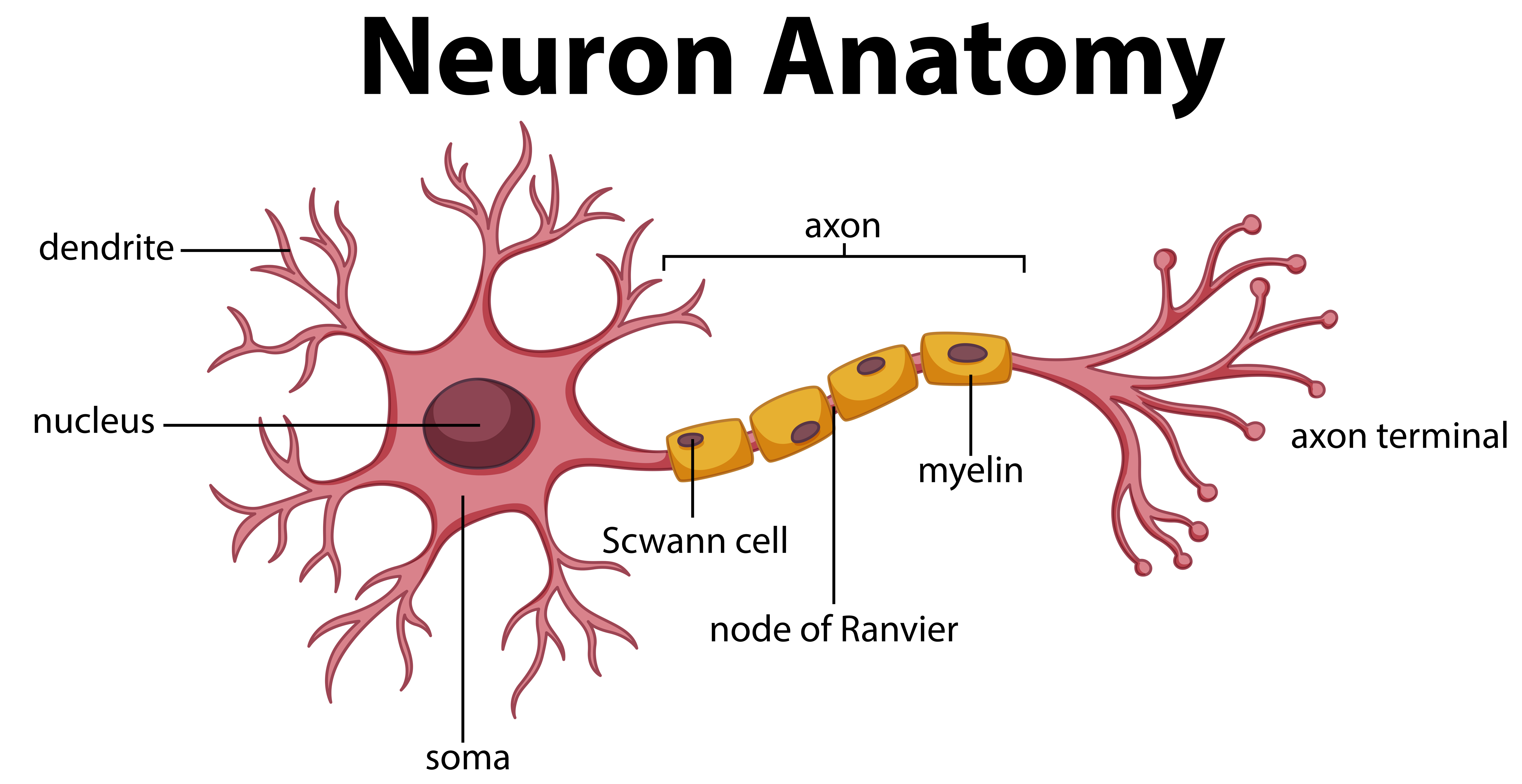
Nervous System Diagrams To Label Teaching Resources | TpT Digitally interactive drag and drop labeling activity to learn the structures of the brain and neuron. This reviews the structure, function and information processing done by the lobes and parts of the brain along with the parts of a nerve cell.
Nervous system drawing with label
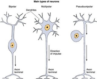
Calcium puts myosin to work (video) | Khan Academy Autonomic vs somatic nervous system. Thermoregulation by muscles. Next lesson . Skeletal system. Current time:0:00Total duration:10:03. 0 energy points. Test prep · MCAT · Organ systems · Muscular system. Calcium puts myosin to work. Google Classroom Facebook Twitter. Email. Muscular system. Practice: Muscular system questions. Myosin and actin. How … Answered: 1) Make a drawing of the nervous system a... |24HA 1) Make a drawing of the nervous system and label the brain, spinal cord, motor nerve and sensory nerve. 2) Provide an example of how the central nervous system and the peripheral nervous system work together to maintain homeostasis. Labeled Neuron Diagram - Science Trends Neurons are a type of cell and are the fundamental constituents of the nervous system and brain. Neurons take in stimuli and convert them to electrical and chemical signals that are sent to our brain. There are 3 major kinds of neurons in the spinal cord: sensory, motor, and interneurons. Neurons communicate vie electrical signals produced by ...
Nervous system drawing with label. Illustration of human nervous system with labels - Shutterstock High usage Superstar Shutterstock customers love this asset! Item ID: 101843320 Illustration of human nervous system with labels Formats EPS 671 × 800 pixels • 2.2 × 2.7 in • DPI 300 • JPG Contributor G GraphicsRF.com Similar images See all Similar video clips Nervous System Structure and Function Labeling and Coloring Activities ... Nervous System Structure and Function Labeling and Coloring Activities - Downloadable Only. Price: $6.95. Add to Cart. View Cart. This bundle includes 14 Nervous System anatomy assessment activities for high school and college anatomy students, including: diagram labeling and coloring pages. All answer keys included. (28 pages total). PharmaCircle This website uses cookies to help provide you with the best possible online experience. Please read our Terms & Conditions and Privacy Policy for information about ... Module 6 nervous system objectives draw and label a Module 6 nervous system objectives draw and label a SchoolWestern University Course TitlePHYS 2130 Type Notes Uploaded Bynpaulose Pages183 Ratings71%(7)5 out of 7 people found this document helpful This previewshows page 50 - 53out of 183pages. Students who viewed this also studied Western University PHYSIOLOGY 2130
Label the Eye Worksheet – Teacher-Made Learning Resources In this resource, you’ll find a 2-page PDF that is easy to download, print out, and use immediately with your class. The first page is a labelling exercise with two diagrams of the human eye. One is a view from the outside, and the other is a more detailed cross-section. On the second page, you’ll find a set of answers showing the properly labelled human eyes, designed to help you … Draw Your Nervous System | AMNH Cut a sheet of paper that is longer than the height of your friend. (Or, tape sheets of paper together.) Place the paper on a hard, smooth floor. Have your friend lay down in the middle of the paper. Use a black marker to draw an outline of your friend's body. Next, draw the different parts of the nervous system. Then, label them. Retina - Wikipedia In vertebrate embryonic development, the retina and the optic nerve originate as outgrowths of the developing brain, specifically the embryonic diencephalon; thus, the retina is considered part of the central nervous system (CNS) and is actually brain tissue. It is the only part of the CNS that can be visualized non-invasively A&P Chapter 11 Nervous System 2 Homework Flashcards - Quizlet The image depicts an example of the autonomic nervous system and an example the somatic motor system. Identify each of the examples. Then, for each label, determine whether it describes the autonomic nervous system or the somatic motor nervous system, and drag it into the appropriate box .
Biology (BIOL) < Johnson County Community College - JCCC VII. Nervous System. A. List the functions of the nervous system. B. Describe how nervous tissue is organized. C. Identify the types of nerve cells. D. List functions of nerve cells. E. Identify structures within nerve cells. F. Explain how an injured nerve may regenerate. G. Explain nerve cell potentials. H. Summarize the events at a synapse. Sympathetic Nervous System Diagram - SmartDraw Sympathetic Nervous System Diagram. Create healthcare diagrams like this example called Sympathetic Nervous System Diagram in minutes with SmartDraw. SmartDraw includes 1000s of professional healthcare and anatomy chart templates that you can modify and make your own. 1/75 EXAMPLES. Art Therapy Exercises To Help Reduce Feelings of Anxiety Art therapy helps facilitate cognitive disruption by moving attention away from rumination. This redirection of attention away from worrying then helps to regulate the nervous system. Art therapy allows us to express ourselves visually and rely less on verbal expression. Verbal expression can be a difficult task if a client is catastrophising ... Calcium puts myosin to work (video) - Khan Academy Now, I'm gonna actually take a further zoom-in. Let's say you actually wanted to zoom in to something like this, this white box. Kind of take a look at what that might look like. Let's see that. I'm gonna make a little bit of space. Let's just keep that scene like that. Let me start my drawing the actin, it's gonna look something like this.
Diagram of Neuron Anatomy - Download Free Vectors, Clipart Graphics ...
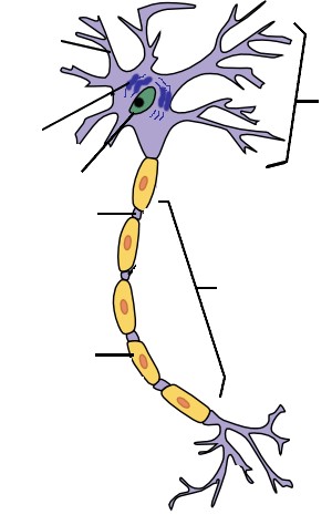
Nervous System - Label the Neuron - TheInspiredInstructor.com Nervous System - Neuron: Nerve Cell. Choose the correct names for the parts of the neuron. This neuron part receives messages from other neurons. This neuron part sends on messages to other neurons. This neuron part gives messages to muscle tissue. This neuron part processes incoming messages. This neuron part contains instructions for making ...
Nervous System Worksheet - WikiEducator
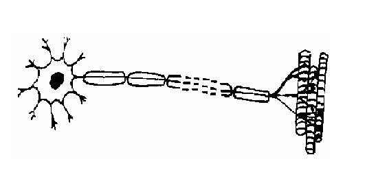
Solved Week 07 Nervous System Laboratory Exercise 21 Nerve | Chegg.com Week 07 Nervous System Laboratory Exercise 21 Nerve Tissue INSTRUCTIONS: 1. Draw the microscopic features of the following neurons and label the identifying/differentiating features. 2. Write the labels on right side 3. Do not abbreviate nor use acronyms. Complete the name of the structures.
Chap 9: The Nervous System (part 1)
Drawing Of The Brain With Labels - Painting Valley label system easy physiology infant coronal neat nervous spinal simple cord rat Brain Diagram Labele... 633x512 41 0 Diagram Of Brain Ste... 525x258 27 2 Brain Clipart Withou... 299x237 16 1 Diagram Of The Human... 232x300 14 1 Draw It Neat How To ... 650x515 9 0 Labeled Fetal Pig Br... 1024x631 9 0 Brain - Drawing Of T... 492x334 5 0

Human Nervous System Medical Vector Illustration Diagram With ...
Retina - Wikipedia Structure Inverted versus non-inverted retina. The vertebrate retina is inverted in the sense that the light sensing cells are in the back of the retina, so that light has to pass through layers of neurons and capillaries before it reaches the rods and cones. The ganglion cells, whose axons form the optic nerve, are at the front of the retina; therefore the optic nerve must cross through …

Labeled Diagram Of The Neuron Stock Vector - Illustration of anatomical ...
Nervous system: Structure, function and diagram | Kenhub The nervous system is a network of neurons whose main feature is to generate, modulate and transmit information between all the different parts of the human body. This property enables many important functions of the nervous system, such as regulation of vital body functions ( heartbeat, breathing, digestion), sensation and body movements.

Nervous System
Nervous System Reading, Color Diagrams, Labeling and Coloring ... This bundle includes 41 Nervous System Anatomy Reading, Color Diagrams, Labeling and Coloring Activities for high school and college anatomy students, including: reading, color diagrams, labeling and coloring (66 pages total). The nervous system is very complex with challenging histology and physiology.
Nervous system - BIOLOGY4ISC
GCSE Biology Question and Answers 2020/2021 - S-cool a) Activity of the nervous system is reduced. Responses slow you down and you become sleepy. (1 mark) b) Provide a feeling of sleepiness and calm when first taken. Motivation decreases and your body deteriorates both mentally and physically. (1 mark) c) Increases activity of the nervous system. Raises alertness, emotions or mood.
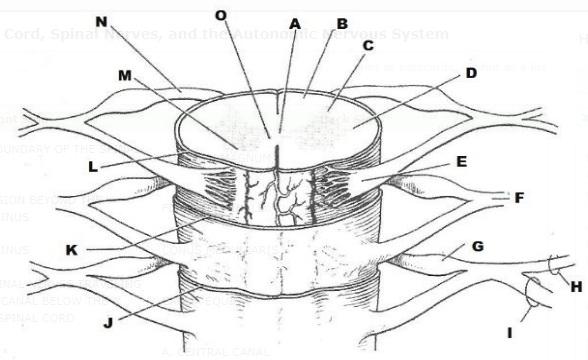
Print Exercise 21: Spinal Cord, Spinal Nerves, and the Autonomic ...
Human Nervous System Structure and Functions Explained With Diagrams The spinal cord is a long tubular structure composed of nervous tissue and support cells. It is around 45 cm long in men and 43 cm long in women. It extends from the brain up to the space between the first and the second lumbar vertebrae. It transmits neural signals between the brain and other body parts.

A Guide to Understand Neuron with Neuron Diagram - Edrawsoft To learn about the structure of the neurons, the students can use a neuron labeled diagram. The students may follow these steps to make their neuron diagram, but the process is complex: 3.1 How to Draw a Neuron Diagram from Sketch Step 1: First, the students need to draw a circle. Based on it, they need to draw a star-like shape.
Post a Comment for "45 nervous system drawing with label"